


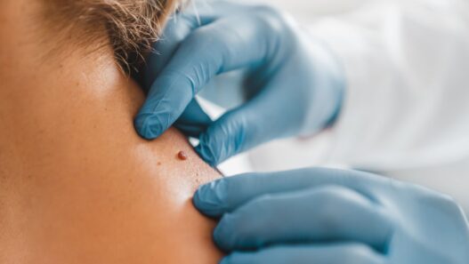


If I’ve learned anything from writing about skin cancer for many years, it’s that it can easily disguise itself as a pimple or common sunspot, and it doesn’t always look threatening. That’s why it’s so important to get an annual skin check, though those with fair skin, a family history or a personal history of skin cancer may need them more often. During the exam, a board-certified dermatologist or physician assistant can examine you from head to toe and flag anything that may need monitoring, treatment or a biopsy—or simply give you peace of mind until your next visit.
Here, we’re giving you a closer look at the different types of skin cancer so you know which changes may warrant a trip to the derm.
Featured Experts
- Deborah Sarnoff, MD, a board-certified dermatologist based in New York
- Orit Markowitz, MD, a board-certified dermatologist based in New York
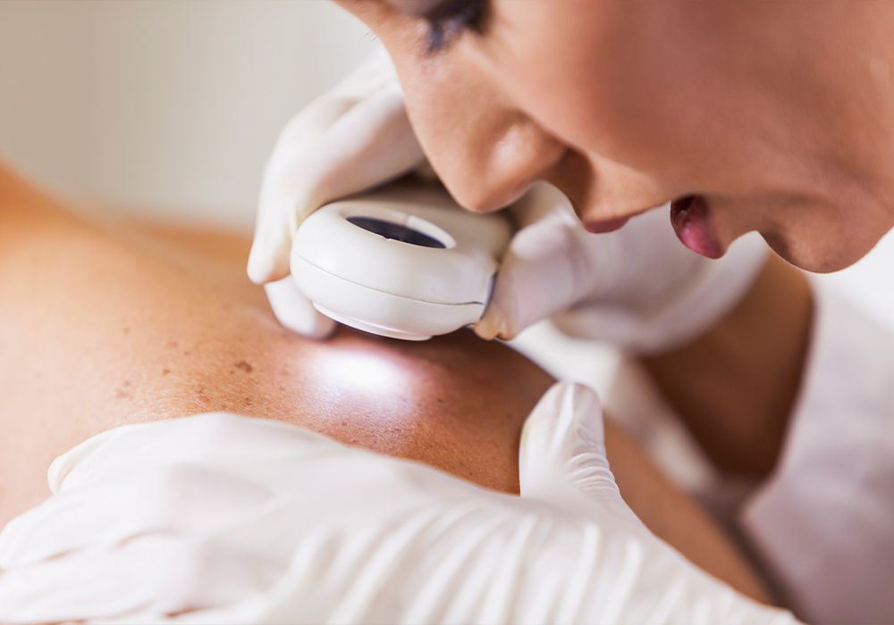
What to Look for During a Skin Check
New York dermatologist Orit Markowitz, MD, says that when it comes to skin cancer, it’s helpful to remember the ABCDEs for moles and spots. Here’s what that means, according to the American Academy of Dermatology:
- A for Asymmetry: Half of the spot is unlike the other half.
- B for Border: The spot has an irregular, scalloped or poorly defined border.
- C for Color: The spot has varying colors from one area to the next, such as shades of tan, brown or black or areas of white, red or blue.
- D for Diameter: Melanomas are usually greater than 6 millimeters, or about the size of a pencil eraser, but when diagnosed, they can be smaller.
- E for Evolving: The spot looks different from the rest or is changing in size, shape or color.
Why Skin Cancer Can Look Different on Everyone
New York dermatologist Deborah Sarnoff, MD says it’s important to remember that skin cancers can look very different from one person to another depending on skin tone, size, type and where they appear on the body. “For example, melanoma is often pigmented tan, brown, black or even blue. But amelanotic melanoma lacks pigment and appears as a skin-tone or pink lesion.”
Ahead, we’re sharing photos of different types of skin cancer, from basal cell carcinoma to melanoma, to show how varied they can appear.
What Skin Cancer Can Look Like
1 / 14

2 / 14

3 / 14

Photo provided by the Skin Cancer Foundation
4 / 14

5 / 14

Photo provided by the Skin Cancer Foundation
6 / 14

7 / 14

Photo provided by the Skin Cancer Foundation
8 / 14

9 / 14

Photo provided by the Skin Cancer Foundation
10 / 14

11 / 14

Photo provided by the Skin Cancer Foundation
12 / 14

13 / 14

Photo provided by the Skin Cancer Foundation
14 / 14